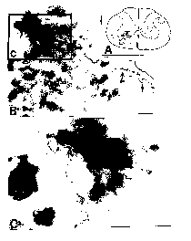
Figure 4. Charting (A) of orthograde (lines and stippling) and retrograde (dots) labeling in the cervical spinal cord, resulting from an injection of BDA made into the right CVLM and an injection of CTb-HRP made into the left diaphragm. Brightfield photomicrographs (B, c, C) of a phrenic motor neuron in the left ventral horn that has been retrogradely labeled by CTb-HRP. B. In addition to the retrogradely labeled phrenic motor neurons, axons and axon varicosities (arrows) orthogradely labeled with BDA can be seen. C. Higher magnification of the labeling seen within the rectangle (c) of B. Note the labeled varicosities (arrows) that are in apparent contact with the phrenic motor neuron. Each scale bar = 50 µm.