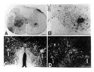
Figure 3. A composite of photomicrographs taken of orthogradely labeled CVLM-spinal axons. A. Brightfield photomicrograph of a spinal cord section taken at mid-cervical levels, in a case (NA-11) where BDA had been injected into the left CVLM. On the left side of the spinal cord, numerous BDA-labeled axons are seen traversing the lateral and ventral funiculi. On the right side, a labeled terminal field can be seen within the ventral horn. B. Higher magnification of the terminal field seen in the right ventral horn of A. Axons contributing to the terminal field can be seen to originate from the lateral and ventral funiculi (medium solid arrows), as well as from the contralateral spinal cord (small paired arrows). C. Darkfield photomicrograph of a spinal cord section taken at mid-cervical levels, in a case (NA-17) where WGA-HRP had been injected into the left CVLM. The resultant terminal field labeling (solid white arrows) is seen bilaterally within the ventral horns. Note that, in addition to the prominent terminal field in the right ventral horn, there is also a terminal field in the left ventral horn. D. A darkfield photomicrograph, also taken from case NA-17, in which the terminal fields (solid white arrows) within each of the ventral horns exhibit nearly equal density. Each scale bar = 300 µm.